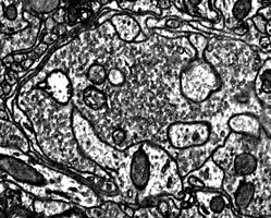
bouton

Description of Dataset
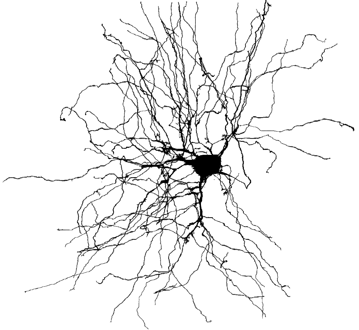

    thalamocortical relay cell
Home
Accessing the Data
Description of the dataset Guide to structures in the dLGN dataset
Project Registry

*The data provided below is currently for publication review purposes only.  At the time of publication, use of the raw data, segmentations and analytical tools will be open to all researchers.


The mouse dorsal lateral geniculate nucleus (dLGN): In mammals, the dLGN is the primary relay between the retina and the visual cortex.  The primary excitatory feed forward pathway thro
ugh the dLGN consists of retinal ganglion cell (RGC) innervating thalamocortical cells (TC) which then project on to the cortex.  The circuit is complicated by a number of factors.  First there are many different kinds of RGC that produce diverse input patterns on thalamocortical cells (see forthcoming publication).  Secondly, RGCs are just one on of many populations innervating the thalamocortical cells. To help identify the various cells and structures of the dLGN, you can refer to this guide while viewing the dLGNs1 data.

The imaged volume: The dLGNs1 volume is a 400 um x 600 um x 280 um wide volume of P32 mouse dorsal lateral geniculate nucleus (dLGN). This volume includes the entire depth of the dLGN. It is positioned latterly, in the monocular region of the dLGN.  The original pixel size of the images is 4 x 4 x 30 nm.  Due to compression during cutting, we estimate the the biologically correct pixel size is 4 x 6 x 30 nm.   The raw dataset consists of approximately 100 trillion voxels (100 TB uncompressed). We have also made available a downsampled version of this dataset in which the voxel size is 16 x 16 x 30 nm.  We find that this resolution is sufficient for many tracing projects.