(206) 601-8008 joshmorgan@fas.harvard.edu Harvard University Dept. Molecular and Cell Biology 52 Oxford St Cambridge MA, 0213 EDUCATION AND TRAINING
1997-2001B.A.
in Neurobiology, New
College of the
University of South Florida, Sarasota,
FL (This university emphasized self guided learning over grades,
hence my GPA =
0)
2001-2007Ph.D.Neuroscience,
Washington
University, St. Louis, MO,
Division of Biology and Biomedical Sciences, Department
of Anatomy & Neurobiology.
Dr. Rachel O.L. Wong, advisor 2007-2010Postdoctoral
Fellow, University
of Washington,
Seattle, Dept. Biological
Structure,Investigating development of bipolar cell to
retinal ganglion
cell connectivity with Dr Rachel O.L.
Wong 2010-2011Postdoctoral
Fellow, Harvard
Medical School,
Boston MA,Dept.
Neurobiology,Reconstructing
mouse
lateral geniculate nucleus using electron microscopy with Jeff Lichtman
and Clay
Reid 2011-Postdoctoral
Fellow, Harvard,
Cambridge MA, Molecular
and
Cellular Biology / Center for Brain Science,Reconstructing mouse lateral geniculate nucleus using
electron
microscopy with Jeff Lichtman TEACHING AND RESEARCH POSITIONS
1999 Curatorial
Assistant, Allen
Museum of Entomology 2000-2001Research
Assistant, New
College of the
University of South Florida,Recorded
telencephalic
visual responses in fish with Leo Demsky 2002Teaching
Assistant,
Neurophysiology Lab,
Washington University
2007 The
Spencer T. and Ann W. Olin Fellow Awarded
to one graduate each year within the Division of Biology and
Biomedical Science
for excellence in biomedical research. 2008Vision
Training Grant NEI-UW.
Stipend
support for postdoctoral researcher within the department of Biological
Structure. 2009Fundamentals
of Neurobiology Training Grant NIH-Harvard
Medical School.
Stipend
support for postdoctoral research within the department of
Neurobiology
2015 - Conte Center Award, Postdoctoral research funding from theNIMH Silvio Conte
Center grant -
Imprinting a connectome: developmental circuit approach to
mental illness 2015Conte Center Award for
Outstanding Outreach & Community Building Awarded for teaching and mentoring
highschool students
through the Conte Center’s community outreach program
PRIMARY RESEARCH ACCOMPLISHMENTS
·In
order to understand the how neurons remodel their synaptic
connectivity during development,
I used three dimensional optical imaging techniques to track the
development of
neural circuits in the retina. I found that some
properties of the
circuit, namely the alignment of visual maps in the inner and
outer retina
(Morgan 2006, Nat. Neuro.)
and the
Gaussian shape of receptive fields (Morgan 2008, Neural Dev.), emerged early in development,
apparently as a
consequence of the geometry of the tissue. Other circuit
properties, such as
the number of synapses formed between pairs of cells, depended
on activity
dependent synaptic remodeling (Kerschensteiner 2009, Nature). In particular, early in
development, two types of
bipolar cells formed synapses on the same type of retinal
ganglion cell at the
same rate. This balance was then upset by an activity dependent
enhancement of
the connectivity of one, but not the other, bipolar cell type
(Morgan 2011, Neuron).
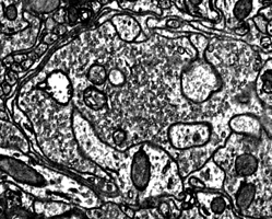
·In
order to map the synaptic connectivity of thousands of neurons
in the same
piece of tissue, I helped develop high throughput electron
microscopy imaging
techniques.This
work included improving
protocols for tissue staining, sectioning and imaging, helping
to write the
software that made automated imaging possible and writing code
for the analysis
of connectivity patterns (Morgan and Hayworth, 2014, Frontiers in Neural Circuits; Kasthuri 2015, Cell). I then used these techniques to produce a
serial section
electron microscopy image volume of unprecedented size (100
trillion
voxels)(submitted).
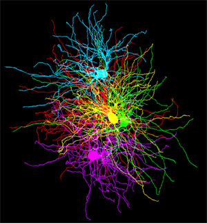
·In
order to understand how axons coordinate their innervation of
target cells, I
mapped the synaptic organization of a large EM volume of mouse
visual thalamus
(submitted). Contrary to the view that visual information
is simply
relayed through the thalamus, I found that retinal inputs
generated complex
networks in which different kinds of retinal axons converged on
the same target
cells. I also found that same retinal axons produced
different synaptic
motifs depending on which target cell they were innervating.
Finally, I found
that sets of axons generated the same input pattern on the
dendrites of
multiple target cells by forming local fascicles that hopped
from one dendrite
to the next.
PUBLICATIONS Martin VV, Beirlein M, Morgan JL, Rothe A,
Gee KR. (2004) Novel
fluo-4 analogs for fluorescent calcium measurements. Cell Calcium.36: 509-14. Morgan J,
Wong R. (2004) Single dendrite seeks stable relationship. Nat Neurosci. 7: 205-6. Lohmann C, Mumm J, Morgan J, Godinho L,
Schroeter E, Stacy
R, Wong WT, Oakley D, Wong ROL. (2005) Live Imaging of the
developing retina.
In: Yuste R, Konnerth A, editors. Imaging In Neuroscience and
Development
171-184 Lohmann C, Demas J, Morgan JL, Wong ROL,
(2005) A Practical
Guide to: Calcium Imaging of the Retina. In: Yuste R, Konnerth
A, editors. Imaging In
Neuroscience and Development Cold
283-288 Mumm JS, Godinho L, Morgan JL, Oakley DM,
Schroeter EH,
Wong RO. (2005) Laminar circuit formation in the vertebrate
retina.Prog.
Brain Res. 147: 155-69 Morgan J,
Huckfeldt R, Wong ROL. (2005) Imaging techniques in retinal
research. Experimental Eye Research 80: 297-306
Morgan JL,
Dhingra A, Vardi N, Wong RO. (2006) Axons and dendrites
originate from neuroepithelial-like
processes
of retinal bipolar cells. Nat.
Neurosci. 9: 85-92 (Highlighted
by
Faculty 1000)
Morgan JL, Wong
RO. (2008) Ballistic labeling with fluorescent dyes and
indicators.Curr
Protoc Neurosci. Chapter 2: Unit 2.11.
Morgan JL,
Schubert T, Wong RO. (2008) Developmental patterning of
glutamatergic synapses
onto retinal ganglion cells. Neural
Develop, 3:8(Highlighted by J. Bio.)
Huckfeldt R.M, Schubert T.*, Morgan J.L.*, Godinho
L. Di Cristo G,
Huang J.Z. and Wong R.O.L (2008)
Transient
neuronal processes regulate spatial distribution of a class of
retinal
interneuron Nature
Neuroscience 12, 35-43
Kerschensteiner D., Morgan J.L., Parker
E.D., Lewis R.M.,
Wong R.O.L(2009)
Neurotransmission
selectively regulates synapse formation in parallel circuits in
vivo.Nature
460: 1016-20.
Williams PR, Morgan JL,
Kerschensteiner D, Wong ROL,
(2011) Live imaging of developing retinal circuits. In: Sharpe
J., Wong R.,
Yuste R., Imaging in
Developmental
Biology: A Laboratory Manual, (Cold Spring Harbor
Laboratory Press)
Morgan JL, Kerschensteiner
D, (2011) Balistic labeling of developing retinaln. In: Sharpe
J., Wong R.,
Yuste R., Imaging in
Developmental
Biology: A Laboratory Manual, (Cold Spring Harbor
Laboratory Press) pp. 177-199.
Morgan J.L.,
Soto F., Wong R.O.L.,Kerschensteiner
D.,
(2011) Development of cell type-specific connectivity patterns
of
converging excitatory axons in the retina. Neuron
71: 1014-21
Schwartz GW, Okawa H, Dunn
FA, Morgan JL,
Kerschensteiner D,
Wong RO, Rieke F., (2012) The spatial structure of a nonlinear
receptive field.
Nat. Neurosci. 15:
1572-80
Morgan JL, Lichtman
JL., (2013) Why not connectomics? Nature
Methods10:
494-500
Morgan
JL*, Hayworth KJ*,
Schalek R, Berger DR, Hildebrand DG, Lichtman JW. (2014) Imaging
ATUM ultrathin
section libraries with WaferMapper: a multi-scale approach to EM
reconstruction
of neural circuits. Front
Neural Circuits. 8:68.
Kasthuri N,
Hayworth KJ, Berger DR, Schalek RL, Conchello JA, Knowles-Barley
S, Lee D,
Vázquez-Reina A, Kaynig V, Jones TR, Roberts M, Morgan JL, Tapia JC, Seung HS, Roncal WG,
Vogelstein JT, Burns R,
Sussman DL, Priebe CE, Pfister H, Lichtman JW., (2015) Saturated
reconstruction
of a volume of neocortex. Cell
162: 648-61
Morgan,
JL and
Lichtman,
JW(in press) Digital tissue. In Cellular Connectomics: Reconstruction of complete
neural wiring
diagrams.Editors:
Brigmann, K and
Helmstaedter, M., Elsevier Press.
Morgan,
JL.,Berger
D.B., Wetzel A.W., Lichtman J.W. (Submitted to Cell) The fuzzy
logic of
network connectivity in mouse visual thalamus.